

Author(s): John A. DeBello, DPM, Kordai I. DeCoteau, DPM, and Eric Beatty, DPM
Publication: Podiatry Today, January 2006
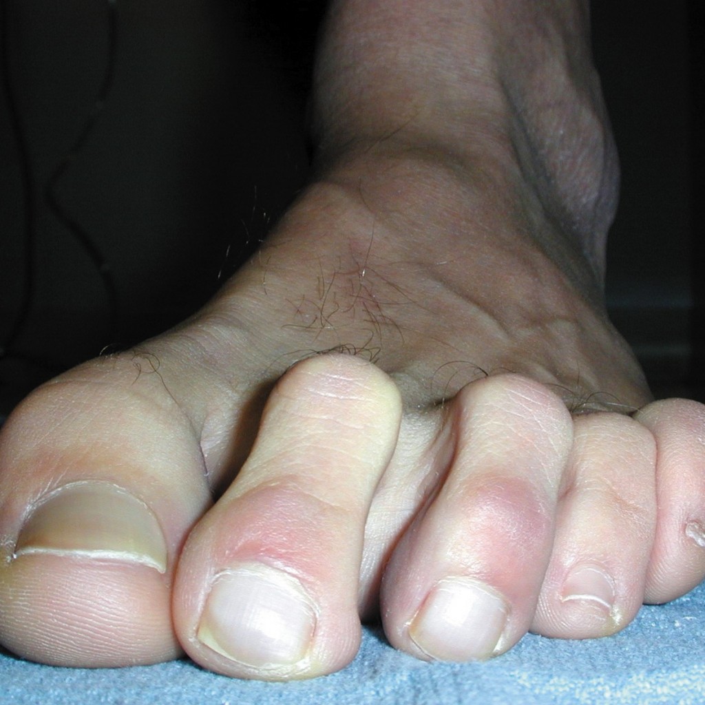
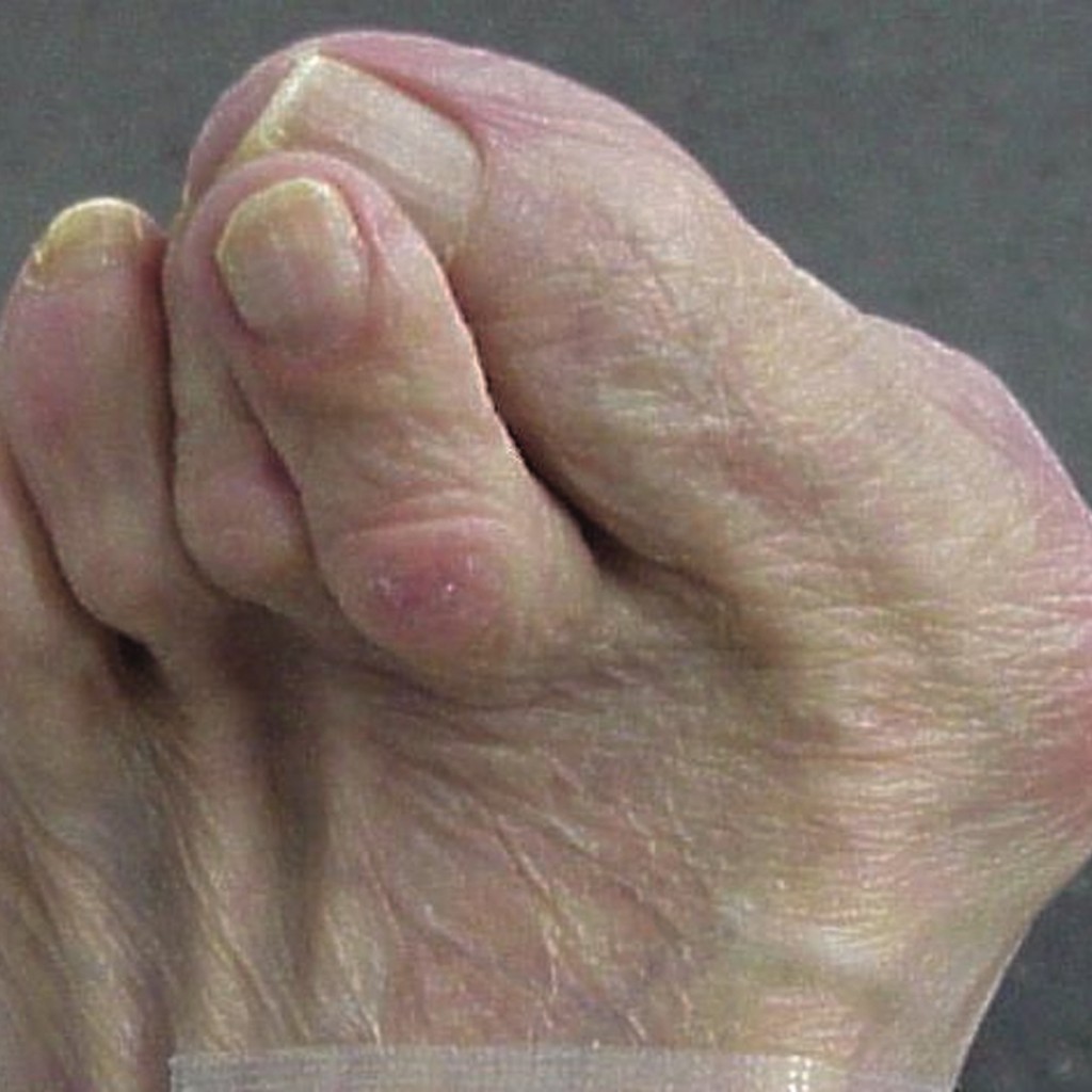
Hammertoes may have an etiology that is either congenital or acquired. Pain and cosmetic appearance are the leading factors for patients wanting surgical intervention for hammertoe deformities. While there are a variety of approaches for hammertoe correction, we have found success with a novel approach that emphasizes the use of medial and lateral incisions. Typically, surgeons use dorsal linear, dorsal longitudinal semi-elliptical, dorsal transverse semi-elliptical, plantar longitudinal and medial/lateral incisions in hammertoe surgery.1However, we feel the medial and lateral incisions provide adequate exposure to achieve the goal of deformity correction and facilitate cosmetic results that patients appreciate and prefer. We typically perform medial incisions to the second and fifth digits, and opt for lateral incisions to the third and fourth digits. Incorporating these incisions addresses the longer aspect of the toe (i.e., medial or lateral) and prevents suture contracture. The procedures involve an arthroplasty or arthrodesis of the proximal interphalangeal joint (PIPJ) while leaving the extensor and flexor tendons intact. One may also perform a flexor tendon transfer depending on the severity of the deformity.
A Step-By-Step Guide To Surgery
When beginning the hammertoe procedure, one should evaluate the involved digit and anesthetize it with 3 to 4 cc of 0.5% bupivicaine in the traditional v-block fashion. Using a number 15 blade, make a 2 cm linear incision just distal to the PIPJ and ending before the web space on the medial/lateral aspect of the digit. Identify all neurovascular structures and retract them as necessary. Deepen the incision with sharp dissection to the
capsule of the PIPJ. One would then identify the extensor mechanism. Pass the blade between the extensor tendons and bone, separating the bone from the extensor hood. Then release the medial and lateral collateral ligaments, allowing access to the head of the proximal phalanx. Using a double action bone cutter, transect the phalangeal head at the surgical neck and remove it in toto.2 Use a bone rasp to smooth and remove any bone spicules. If one has not achieved the correction, the surgeon should proceed to perform an arthrodesis. Using a 0.045-inch Kirschner wire, drive the wire retrograde through the medullary canal of the middle phalanx, exiting through the distal tip of the digit. One would then drive the Kirschner wire anterograde through the distal and middle phalange, and through the PIPJ.2 If the digit is subluxed, perform a flexor tendon transfer. Identify the flexor tendon and release the fully intact tendon from its capsular attachments. Transect the tendon as far distally as possible through the original incision. Split the tendon longitudinally in half and reapproximate it dorsally over the proximal phalanx.3,4 One can extend the incision proximally to allow for a metatarsophalangeal joint release without transecting either tendon. The surgeon can transect the collateral ligaments. Using a McGlamry elevator, one can free the metatarsal head dorsally. Irrigate the surgical site with normal saline. If you have performed a flexor tendon transfer, you should reapproximate with 3.0 Vicryl. Close the skin using 4.0 nylon in a simple suture fashion. Dress the toe with a non-adherent dressing and use a Betadine sterile bandage to help splint the toe. Have the patient wear a postoperative shoe. Remove the sutures within 10 to 14 days and remove the Kirschner wire after four weeks.
How The Procedure Offers Key Benefits
The success of these procedures is due to the location of our incision and the minimal contact/pressure of the incision site. Neurovascular insult is always a concern. However, one can make the incision parallel to the neurovascular structures in order to minimize the risk of any trauma that could occur. The location of our incision also provides adequate exposure to the structures for surgical correction. This helps simplify the technique. The procedure spares tendons the risk of accidental transection that can occur more frequently through more traditional incisions. There is also increased postoperative stability of the digit with minimal trauma to the periarticular structures.2 Another benefit of the procedure is that patients do not bear weight on the surgical site. This is particularly ideal for fifth digit procedures in preventing scarring, pain and possible dehiscence. The procedure also reduces a possible flail toe as the surgeon would not transect the extensor tendon. Additionally, one may correct a mallet toe in the same fashion as a hammertoe by extending the incision distally for exposure. Finally, the cosmetic result lends itself to higher numbers of satisfied patients.
In Conclusion
With plastic surgery techniques becoming popular in foot surgery, we believe that the medial/lateral technique is a superior approach to digital surgery. It is relatively easy in terms of technical difficulty and a surgeon can perform it within the same amount of time as a traditional incision. The medial incision increases the chance that patient is more apt to have appropriate surgical management. Dr. DeBello is a Fellow of the American College of Foot and Ankle Surgeons. He is a Diplomate of the American Board of Podiatric Surgery and is board certified in foot surgery. He is affiliated with the Podiatric Surgical Department at the Our Lady of Mercy Hospital in Bronx, N.Y. Dr. DeCoteau is affiliated with the Podiatric Surgical Department at the Our Lady of Mercy Hospital at Bronx, N.Y. Dr. Beatty is affiliated with the Podiatric Surgical Department of Our Lady of Mercy Hospital in Bronx, N.Y. Dr. Burks is a Fellow of the American College of Foot and Ankle Surgeons, and is board certified in foot and ankle surgery. Dr. Burks practices in Little Rock, Ark. Editor’s Note: For related articles, see “How To Handle Complications Of Hammertoe Surgery” (page 36, September 2005 issue) or “The Top Eleven Pearls For Hammertoe Surgery” (page 24, April 2002 issue). Also visit the archives at www.podiatrytoday.com.
References:
- Mozena JD, Yeske M, Jones PC. Hammertoe surgery and the medial incisional approach. Podiatry Today. December:50-53, 1998.
- Ohm OW, McDonnel M, Vetter WA. Digital Arthrodesis: An Alternate Method for Correction of Hammer Toe Deformity. Journal of Foot Surgery. 29(3):207-211, 1990.
- Gazdag A, Cracchiolo A. Surgical Treatment of Patients with Painful Instability of the Second Metatarsophalangeal Joint. Foot and Ankle Int. 19(3):137-143, 1998.
- Mendicino RW, Statler TK, Saltrick KR, Catanzariti AR. Predislocation Syndrome: A Review and Retrospective Analysis of Eight Patients. J Foot Ankle Surg. 40(4):214-224, 2001.
– See more at: http://www.podiatrytoday.com/article/5023#sthash.ouSaPyWU.dpuf